A válvula de uretra posterior é uma malformação caracterizada por uma membrana obstrutiva na uretra afetando o fluxo urinário, o que pode causar insuficiência renal.
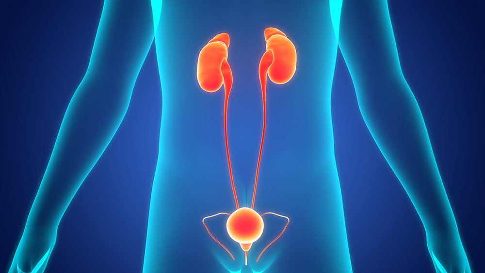
A válvula de uretra posterior (VUP) é uma condição rara que afeta o trato urinário masculino. Essa condição é caracterizada por uma membrana obstrutiva que se localiza na parte de trás da uretra próximo à bexiga, entre a próstata e o esfíncter uretral externo.
De acordo com o médico urologista Dr. Ubirajara Barroso Jr., chefe da Unidade do Sistema Urinário do Hospital Universitário Professor Edgard Santos (UFBA), a incidência de válvula de uretra posterior é em média de 1 para cada 8 mil nascidos vivos.
A válvula de uretra posterior é uma condição que requer compreensão detalhada, diagnóstico precoce e tratamento adequado para evitar complicações graves.
Neste guia, abordaremos o que é, quais são as possíveis causas, sintomas característicos e as opções de tratamento disponíveis para a válvula de uretra posterior. Leia e confira.
O que é válvula de uretra posterior
A válvula de uretra posterior é uma anomalia congênita na estrutura da uretra masculina. Ela envolve uma membrana anormal no interior da uretra, que pode obstruir parcial ou completamente o fluxo urinário. Essa obstrução pode levar a complicações graves se não for tratada adequadamente.
“Na verdade, o que existe não é uma válvula verdadeira, e sim uma membrana, porém como esta nomenclatura, dada no início do século passado, já foi utilizada pelo tempo, ela permanece até hoje”, explica Dr. Ubirajara Barroso, que também é chefe da divisão de cirurgia urológica reconstrutora e urologia pediátrica do Hospital da Universidade Federal da Bahia.
Ele explica que a válvula de uretra posterior pode ser tipo 1, tipo 2 ou tipo 3.
Válvula de uretra posterior tipo 1:
O tipo 1 é o mais comum, representando aproximadamente 95% dos casos. Nessa condição, há a presença de uma membrana que forma um arco aberto na porção inferior da uretra.
“Alguns teorizam que a tipo 1 seria na verdade mais aberta em virtude da sondagem uretral em que todas as válvulas seriam de alguma maneira concêntrica”, explica.
Válvula de uretra posterior tipo 2:
O tipo 2 é caracterizado por uma dobra na mucosa. Segundo Dr. Barroso, alguns especialistas acreditam não ser uma válvula de fato.
Válvula de uretra posterior tipo 3:
O tipo 3 é semelhante a um orifício, sendo uma membrana concêntrica, e é considerado o mais problemático, pois pode causar complicações significativas na bexiga.
“A válvula de uretra posterior causa uma lesão importante na bexiga porque o músculo da bexiga, que é o músculo detrusor, para expelir a urina precisa contrair com uma força cada vez maior, isso vai fazendo com que haja uma hipertrofia muscular e deposição de colágeno, fibrose e aumento da pressão dentro da bexiga”, descreve Dr. Barroso, que destaca que essas contrações podem causar lesão irreversível aos rins, ou seja, uma insuficiência renal.
Quais as causas da válvula de uretra posterior?
A causa exata da válvula de uretra posterior ainda não é totalmente compreendida, mas acredita-se que fatores genéticos e anormalidades no desenvolvimento da uretra durante a gestação estejam envolvidos.
“Estima-se que haja alguma questão genética envolvida, mas como está na uretra posterior, numa região onde está a saída da próstata em uma estrutura chamada verumontanum, acredita-se que seja na formação dessa porção com uma membrana persistente ou na inserção do verumontanum na uretra que está a origem do problema”, relata o médico que também atua com urologia pediátrica e reconstrutora em São Paulo.
Diagnóstico e sintomas de válvula de uretra posterior
Segundo Dr. Barroso, atualmente, a maioria das válvulas de uretra posterior é identificada durante o período pré-natal.
A detecção é realizada quando há:
- Presença de dilatação renal bilateral;
- Dilatação ureteral;
- Bexiga distendida, que não consegue esvaziar adequadamente, apresentando paredes espessas e grossas.
Mais tarde, depois que a criança nasce ou são maiores, a válvula de uretra posterior pode ser diagnosticada por:
- Infecção urinária, geralmente com febre;
- Dificuldade para urinar;
- Demora para iniciar o jato;
- Jato fraco;
- Urgência miccional;
- Incontinência urinária.
O diagnóstico geralmente é feito através de cistouretrografia miccional, um exame por onde é passada uma sonda pela uretra e injetado iodo. O contraste permite visualizar a uretra durante a micção e identificar a presença da membrana obstrutiva.
“A criança tem que estar em posição oblíqua ou de perfil para melhor avaliação de toda a anatomia uretral na busca da presença dessa membrana obstrutiva”, explica.
Tratamento e novas abordagens para válvula de uretra posterior
Dr. Ubirajara Barroso destaca que a válvula de uretra posterior é tratada por meio de endoscopia com a visualização da válvula, onde é realizada uma ablação.
“Para aquelas crianças com insuficiência renal, algumas vezes é necessário fazer uma derivação urinária, que é um desvio de trânsito para a pele de forma temporária, que pode ser da bexiga para a pele, chamada vesicostomia, ou do ureter para a pele, denominada ureterostomia cutânea”, diz.
De acordo com Dr. Ubirajara Barroso Jr., novas abordagens estão sendo exploradas para oferecer tratamentos mais precisos e eficazes. “Uma novidade no tratamento é a sondagem da bexiga à noite, a criança com válvula de uretra tem um cateterismo noturno aberto, essa foi uma ideia do Stephen Koff”, destaca.
Esse método envolve o cateterismo noturno aberto em crianças com válvula de uretra, aproveitando o período em que elas tendem a ter poliúria (produção e eliminação excessiva de urina), garantindo que a bexiga permaneça em baixa pressão por 8 a 10 horas durante o sono. Essa prática tem se mostrado eficaz na redução da incidência de insuficiência renal, proporcionando alívio às bexigas que sofrem com a obstrução da válvula.
Outra inovação importante é o tratamento da válvula de uretra posterior por meio de laser. “Ainda não está muito bem documentada a vantagem desse tipo de tratamento, porém tecnicamente é possível uma ablação da válvula com maior precisão e provavelmente diminuindo os riscos de lesão esfincteriana”, aponta.
Além disso, foi estabelecida a importância do estudo urodinâmico após a ablação da válvula. Esse procedimento é fundamental para avaliar a função da bexiga e iniciar o tratamento precoce de possíveis disfunções, seja por meio do uso de anticolinérgicos ou alfa-bloqueadores.
Cuidados pós-tratamento e acompanhamento
Dr. Barroso enfatiza que mesmo após o tratamento da válvula, estudos urodinâmicos mostram que 75% das pessoas após ablação têm contrações involuntárias da bexiga, o que faz com que a bexiga continue a ser afetada pelo esforço urinário, resultando em alterações persistentes.
“Isso pode levar a sintomas de urgência miccional, risco de incontinência urinária e execução de manobras de contenção para evitar essa perda”, destaca.
O especialista afirma que nesses casos, mesmo que a válvula seja tratada precocemente, a falta de tratamento subsequente da bexiga pode resultar em complicações tardias, incluindo a insuficiência renal.
Além disso, após o tratamento da válvula, devido aos efeitos que ela causa nos rins, ocorre uma alteração na concentração urinária de eletrólitos, resultando em um aumento na produção de urina durante o dia, o que é chamado de poliúria.
“A poliúria pode fazer com que aumente ainda mais a sobrecarga da bexiga e o risco de disfunção da bexiga permanente”, alerta.
É de extrema importância que as crianças, especialmente após o período de desfralde, que tenham passado por ablação da válvula recebam acompanhamento de um urologista para evitar essas alterações no longo prazo e garantir uma qualidade de vida adequada.
Você percebe algum dos sintomas descritos neste texto em seu filho? Ele já passou por tratamento para válvula de uretra posterior? Se sim, é importante agendar uma consulta médica para uma avaliação adequada e garantir que ele receba o acompanhamento necessário para sua saúde urinária.
LEIA MAIS:
Disfunção do trato urinário inferior (DTUI) – entenda o que é